How to Find the Right Working Length with an Apex Locator (and Confirm It on a Radiograph)
Apex locator to APEX, withdraw to 0.5, confirm on an undistorted periapical. A student guide to working length that stops at the constriction.
Every root canal you will ever treat comes down to one number: how far down the canal you work. Get that number right and everything downstream — cleaning, shaping, obturation — has a chance. Get it wrong by a millimetre and you are either leaving infection behind or pushing instruments into the periapical tissues. Here is how to measure it properly, the way current guidance actually asks you to.
Where the canal really ends (hint: not at the tip)
The first mental correction most students need is this: the canal does not end where the root ends. Travelling apically, the canal narrows to its tightest point — the apical constriction — passes the cemento-dentinal junction, opens at the apical foramen, and only then do you reach the radiographic apex you see on the film. Classic anatomical work (Kuttler, 1955) places that constriction roughly 0.5–1 mm short of the radiographic apex.
Working length is defined from a stable coronal reference point — a cusp tip, an incisal edge — down to that apical endpoint. Your target is the constriction, not the foramen and definitely not the radiographic apex. If you have been eyeballing "1 mm short of the apex" and calling it done, you have the right instinct but only half the method.
Why half a millimetre decides the case
Think about what happens at each error direction. Stop short of the constriction and you under-prepare: infected pulp tissue and debris stay in the apical canal, the obturation seals over them, and the lesion never resolves. Go past the foramen and you over-instrument: you traumatise the periapex, invite post-operative pain and flare-ups, and delay healing.
That is why working length is not a box you tick once during access. It is the number that governs every file you place, every irrigation depth, and the final fill. Treat it with the same seriousness you treat your diagnosis.
The gold standard: locator first, film to confirm
Both AAE- and ESE-aligned guidance converge on the same protocol: take an electronic working length with an apex locator, then verify it with an undistorted periapical radiograph.
Why both? Because each method fails in a different way. The electronic apex locator (EAL) measures impedance between a lip clip and a file clip, so it reads the position of the file tip relative to the foramen — but it tells you nothing about canal curvature, root anatomy, or which canal you are actually in on a multi-rooted tooth. The radiograph shows you the tip against the anatomy — but an elongated or foreshortened image will lie to you about length. Used together, they cover each other's blind spots, and the combination is the most reliable working length you can get.
The protocol, step by step
Estimate before you measure. Start from a well-angulated diagnostic periapical and read off an estimated canal length, roughly a millimetre short of the radiographic apex. This is not your working length — it is a planning number that lets you pick sensible file lengths and stops you from over-inserting on the first negotiation.
Set up for a clean reading. Rubber dam on. Pulp chamber fully unroofed. Irrigate and clear gross debris, then suction out the excess — a chamber flooded with conductive fluid is the single most common cause of erratic readings. Choose your coronal reference point deliberately: a sound cusp, an incisal edge, or a surface you have flattened, and set the silicone stopper against it.
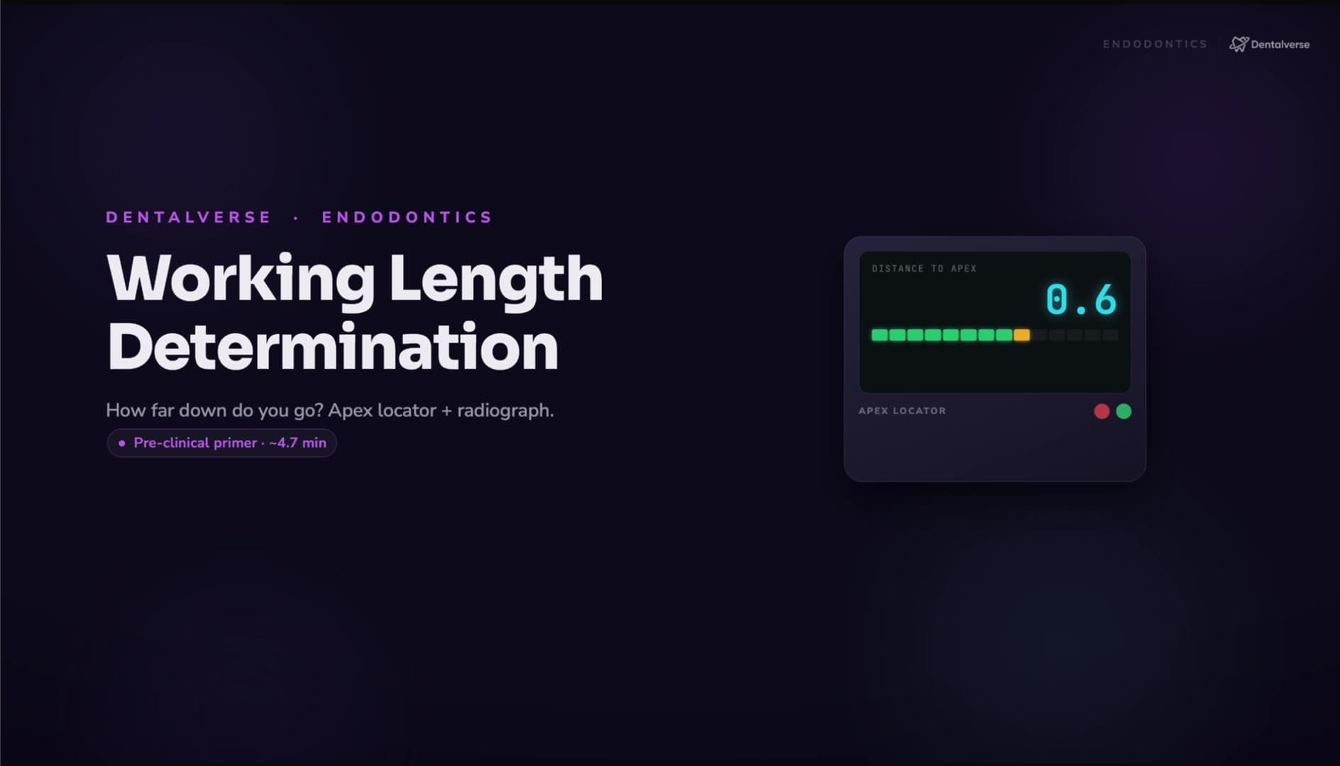
Take the electronic length. Pick a small hand file — a #10 or #15 is typical — attach the file clip, and check the lip clip has good mucosal contact. Advance slowly. The display climbs as the tip approaches the foramen; when it reads zero — APEX — your file tip is at the foramen. That zero is a landmark, not a destination. Withdraw slightly until the display sits around the 0.5 constriction reading: that is your working length. Fix the stopper at the reference point and record the measurement. If the numbers jump around or disagree badly with your radiographic estimate, do not force the file apically — troubleshoot first, and let the radiograph arbitrate.
Confirm on film. With the same file at the electronic length, stopper still at the reference, take an undistorted periapical — paralleling technique preferred. You want to see the file tip sitting about 0.5–1 mm short of the radiographic apex, consistent with the anatomy. If it does not match, Ingle's proportion formula corrects the measurement: true working length = file length × radiographic tooth length ÷ radiographic file length.
When the numbers don't make sense
Erratic readings almost always trace back to electricity, not anatomy. Run this checklist: lip or file clip losing contact — reattach and dry the contacts; chamber flooded with irrigant — suction it; file too small for a wide canal — step up a size; a metal restoration touching the clamp or clip — isolate the contact point.
If the locator shouts APEX while your file is obviously short of length, think mechanical obstruction: a blockage, a ledge, transportation, or calcification. Recapitulate, irrigate, negotiate with small files, and re-establish the glide path before trusting another reading. If it reads long — beyond the apex — suspect a perforation or an open apex: stop, reassess the anatomy radiographically, and reserve CBCT for cases where it is genuinely clinically justified.
Measure again, then write it down
A working length recorded once is not valid for the whole case. Re-check it whenever the coronal reference changes (you flattened a cusp, reduced an incisal edge), whenever you hit a blockage or create a ledge, when you transition from hand scouting to rotary shaping after coronal enlargement, and any time you suspect resorption, perforation, or an open apex.
Then document everything: the reference point, the electronic value and the device used, that a working-length film was taken and verified, and any anomalies — for instance, that the reading was unstable until the chamber was dried. An undocumented length might as well not exist.
Key takeaways
- Aim for the apical constriction, roughly 0.5–1 mm short of the radiographic apex — not the foramen, not the tip.
- Follow the AAE/ESE-aligned sequence: electronic apex locator first, undistorted periapical radiograph to confirm.
- On the locator, advance to the APEX (zero) reading, then withdraw to the 0.5 constriction reading — that is your length.
- Erratic readings are usually electrical (clips, flooded chamber, undersized file, metal contact); premature APEX readings suggest a ledge or blockage.
- Re-check the length whenever the reference point or canal path changes, and chart every measurement.
Study this properly
Review the full step-by-step protocol, instruments, and pitfalls on the Working Length Determination (Apex Locator + Radiograph) reference page. The complete narrated video lesson — with the visual walkthrough of the locator readings and the confirmation film — is inside Dentalverse (start free).
This article is a study aid for dental students, not medical advice — always follow your institution's clinical protocols and your supervisor's guidance.