Dentalverse
Dentalverse
Explore Features
Tooth Atlas
52 teeth mapped
Drug Reference
27 medications
Anesthesia Guide
11 anesthetic agents
Clinical Procedures
135 step-by-step
Tomorrow's Clinic
Prep sheets & day planner
Medical Conditions
83 conditions
Clinical Thinking
40 case simulations
Clinical Mistakes
105 common errors
Video Library
Curated by specialty
INBDE Prep
3,386 study cards
AI Study Tools
Teach Me, Quiz, Chat
View all features
LibraryPodcastBlogPricingFAQLog inTry Free
All articles
Endodonticssurgical endodonticsroot-end fillingMTA

How to Do a Retrograde Root-End Prep and Filling: The 3 mm Seal That Decides Your Apicoectomy

The retro-prep and root-end fill decide whether apical surgery succeeds. Learn the 3 mm geometry rules, the 5-step protocol, and why bioceramics win.

D
Dr. Saleh Albakri
July 13, 2026
3 min read

You can raise a beautiful flap, cut a clean osteotomy, and resect the apex like a textbook — and the case will still fail if the last three millimetres leak. The retrograde preparation and root-end filling are where apical surgery is actually won or lost. Everything before this step is access; this step is the cure.

Why the last 3 mm decide the case

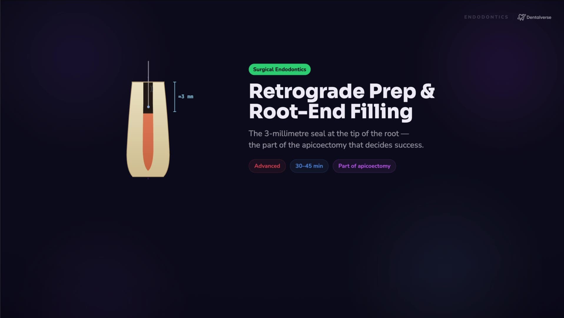
Orthograde endodontics seals the canal system from the crown down. Root-end surgery flips the direction: after the apicoectomy exposes and resects the root tip, you cut a small cavity into the fresh cut face and seal the canal system from the apical side. Two jobs happen at once: the resection removes infected apical anatomy; the retro-prep and fill keep what remains sealed.

The mental model is geometry. A good preparation is about 3 mm deep, centered within the root, parallel to the long axis, and conservative enough to preserve the remaining dentin. Picture a dashed line down the root's long axis — your cavity tracks that line. Every classic failure mode maps back to breaking one of these rules: a shallow prep compromises the seal, an angled prep thins one wall dangerously, and an off-center prep on a curved or thin root risks a strip perforation.

That geometry is why modern technique is defined by ultrasonic retro-tips under the microscope: the tip cuts coaxially, straight down the canal, which a bur can't do without angling. It's also why calcium-silicate bioceramics — MTA-class cements and putties — own the fill: they're biocompatible and, crucially, they set and seal despite blood and moisture — the surgical reality however good your hemostasis. And it's why you read the cut face first — miss an isthmus connecting two canals and you've left the reinfection route wide open.

Key takeaways

  • The retro-prep and fill are the therapeutic core of apical surgery — the resection is only access; the apical seal is the cure.
  • Geometry governs everything: roughly 3 mm deep, centered, parallel to the long axis, preserving dentin — every classic failure is one of those rules broken.
  • Materials are chosen for the field they work in: MTA-class and bioceramic materials took over because they seal in blood and moisture.

Learn the full protocol

The complete walkthrough — the narrated video lesson, the step-by-step protocol with armamentarium and pitfalls, and the night-before prep sheet — is inside Dentalverse. Start with [the reference page](/explore/procedures/retrograde-preparation-and-root-end-filling), then [start free](/signup) to unlock the full lesson.

This article is a study aid for dental students, not medical advice — always follow your institution's clinical protocols.

Inside the app

Put this into practice inside Dentalverse

Every concept in this article is backed by interactive reference material, AI tools, and practice questions.

Tooth Atlas
Tooth #14
Maxillary Left 1st Molar
Roots3 (MB, DB, P)
Canals3–4 (MB2 common)
InnervationPSA nerve
BlockPSA injection
Tooth Atlas→
Drug Reference
AntibioticPenicillin
Amoxicillin
First-line for odontogenic infections
Adult dose
500 mg PO every 8 hours
Prophylaxis (AHA)
2 g PO, 30–60 min pre-op
Contraindication
Penicillin allergy
Drug Reference→
Clinical Case
HR 80
45 min
$520
Turn 2 of 4
A 62-year-old presents with severe pain on tooth #30. BP 148/92. Takes warfarin for AFib.
Next step
Order INR before extraction
Extract immediately
Refer to physician
Clinical Cases→
INBDE Prep
PharmacologyCard 47 / 120
Question
What is the max recommended dose of 2% lidocaine with 1:100,000 epi in a healthy adult?
A3.0 mg/kg
B4.4 mg/kg
C7.0 mg/kg
D10 mg/kg
INBDE Prep→
Procedures
Operative
Class II Composite
8 steps · 30-45 min
1
Anesthesia + isolation
2
Caries removal
3
Matrix + wedge placement
4
Etch + bond
5
Incremental fill
Procedures→
AI Study Chat
When do I prescribe antibiotic prophylaxis for a prosthetic joint patient?
Dentalverse AI
Per the 2015 ADA guideline, routine antibiotic prophylaxis is generally not recommended for prosthetic joint patients. Consult the orthopedic surgeon for high-risk individuals.
ADA 2015JADA
AI Chat→

Continue reading

Ectopic Eruption of the First Permanent Molar: When to Observe, Intercept, or Refer

3 min read

Why You Never Replant a Primary Tooth (and Always Try to Save a Permanent One)

6 min read

How to Extract a Primary Tooth Safely — and Stop the Bleeding Before the Child Goes Home

3 min read

Ready to study smarter?

Sign up free and start studying smarter.

Try Free
Dentalverse

Your dental school companion from Day 1 through graduation. Built for dental students.

Features
Tooth AtlasDrug ReferenceAnesthesia GuideClinical ProceduresTomorrow's ClinicMedical ConditionsClinical ThinkingClinical MistakesVideo LibraryINBDE PrepAI Study Tools
Resources
Explore the LibraryPodcastBlogFAQContact Us
Legal
Privacy PolicyTerms of ServiceRefund PolicyCookie PolicyDisclaimerAI Use PolicyCommunity GuidelinesCopyright

© 2026 Denverse Ltd (Company No. 17146294). All rights reserved.

Educational platform only. Content is not medical or dental advice.