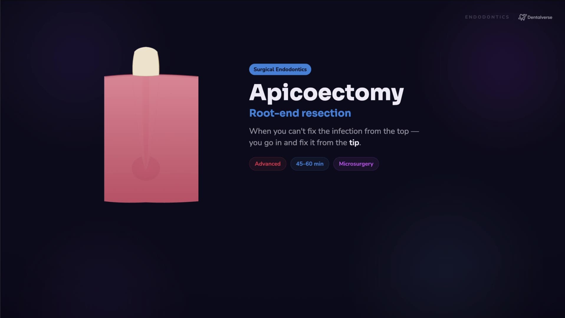
Apicoectomy Step by Step: Why You Resect 3 mm and Cut the Root End Flat
Why apicoectomy resects exactly 3 mm at a 0–10° bevel, the 8-step microsurgery workflow, and the isthmus mistake that makes lesions come back.
Every endodontic failure you'll see on a radiograph has one root cause: somewhere, bacteria still have a pathway. When the coronal route can't reach that pathway — a post you can't remove, a canal you can't negotiate — the answer isn't to give up on the tooth. It's to flip the whole problem and seal the canal system from the other end.
The mental model: seal from the other end
An apicoectomy (root-end resection) is a microsurgical procedure built on one conceptual flip: conventional endodontics seals the canal from the coronal direction; apicoectomy seals it from the apical direction. Same goal — entomb the microbes — opposite approach vector. You access the apex surgically, remove the diseased periapical tissue, cut away the apical portion of the root, and seal from that end.
The selection rule comes before any technique question: is the tooth worth saving, and safely accessible? No microsurgical skill rescues a cracked, non-restorable tooth, and no lesion justifies operating blind next to the mental foramen. Surgery is an advanced option for when retreatment isn't feasible or has failed — not a first move.
The two numbers in the title carry the biology. You resect approximately 3 mm of the apex because that apical 3 mm concentrates the apical ramifications and lateral canals — the leakage pathways that keep the tooth infected. And you cut with a minimal bevel, about 0–10°, because a flat cut improves the seal and exposes fewer dentinal tubules; the old 45°-plus bevels opened huge tubule fields and made sealing unreliable. Cut where the anatomy hides, and cut it flat.
What made all of this predictable is visibility. The operating microscope, ultrasonic root-end preparation, and bioceramic filling materials rewrote the outcomes — largely by letting you inspect the cut root face for the isthmuses, missed canals, and cracks that quietly cause failures.
Key takeaways
- Selection before technique: operate only on a restorable, safely accessible tooth after the coronal route is blocked or has failed.
- Apicoectomy is the mirror image of conventional endodontics — the canal gets sealed from the apical side.
- Microscope, ultrasonics, and bioceramics turned a last resort into a predictable, tooth-saving procedure.
Learn the full protocol
The complete walkthrough lives in Dentalverse: the narrated video lesson, the step-by-step protocol with armamentarium and pitfalls, and the night-before prep sheet. Start from the reference page and start free to unlock the full lesson.
This article is a study aid for dental students, not medical advice — always follow your institution's clinical protocols and your supervisor's guidance.